
The milk-carrying ducts inside your working breast
A small number of main milk ducts open out onto the surface of your nipple
Your milk ducts are an integral part of your breasts' glandular tissue, gathering up your milk and carrying it out through your nipple.
-
Using live ultrasound imaging during breastfeeding, Professor Geddes and her team found that there are sometimes as few as four, sometimes as many as eighteen, main ducts opening onto the face of the nipple. (The average number is nine.) Normality is a very diverse human condition!
-
These main ducts running through the nipple are about two millimetres in diameter at rest, although some main ducts might be just 0.6 millimetres and others up to 4.4 millimetres.
-
Each main duct first branches at about seven or eight millimetres underneath the base of the nipple, into two or three smaller branches which are about 1.3 mm wide. But Professor Geddes and her team sometimes observed ducts even 10 millimetres wide in their resting state inside the breast. Duct widths vary remarkably between lactating women.
Your milk ducts follow densely tangled, profusely branching, and highly irregular paths into the breast
From there, your milk ducts follow densely tangled, intertwined, and highly irregular paths into your breast, "much like the roots of a tree". In this finely interlaced profusion, ducts branch down smaller and smaller within a surprisingly short distance under the nipple, ending in the tiny ductules which drain each lobule (or cluster of alveoli).
-
Your larger milk ducts (and also their accompanying glandular tissue) are located in a 30mm radius around the base of your nipple. The location of most of the glandular tissue just a short distance from the base of nipple means that resistance by the ducts to milk flow is minimised.
-
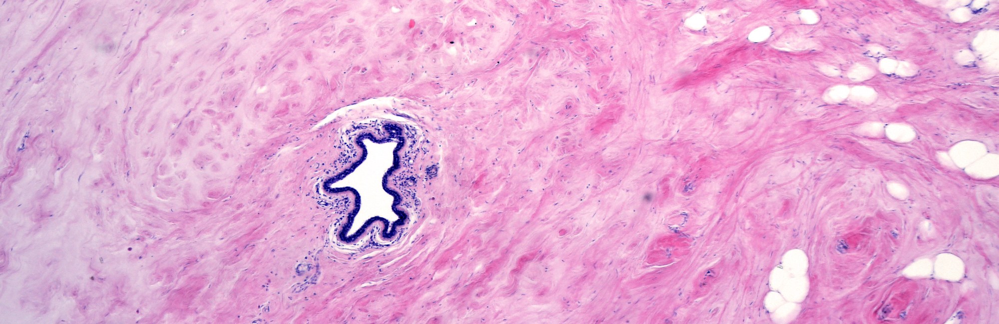
Milk ducts are formed by two epithelial cell layers.
-
The first, facing into the lumen, is a layer of cube-shaped epithelial cells.
-
The second is a layer of star-shaped myoepithelial cells, which contract when flooded with oxytocin. However, this contraction during letdown results in the ducts opening up and shortening.
-
The myoepithelial cells are wrapped in a dense basement membrane.
-
-
Many milk ducts are collapsed at any one time, even though some milk passes into and rests in the ducts as it accumulates in the glands.
-
There are usually more ducts and glandular activity in the lower outer parts of your breast.
Your milk ducts can be easily compressed
Given what we know about milk ducts, above, you can see how milk ducts travel just a millimetre or two deep to the fascia which lies under the skin of the areola and breast. (There is no subcutaneous fat under the nipple-areola complex.) As a result, milk ducts are highly compressible to even to very light surface pressure, occluding in the same way that the lightest touch compresses the veins on the back of your hand.
This is how hand expressing your milk works (although not all women find they can hand express). When there is no vacuum drawing out the milk, positive pressure applied by your fingers can still empty the milk glands and press milk out through the ducts. After a while, the sensory stimulation of hand expressing might trigger a let-down, which helps you because the let-down applies positive pressure to the milk inside your alveoli.
Milk ducts don't have lactiferous sinuses
Professor Geddes was the first to find that we don't have any ‘lactiferous sinuses’ which store your milk, the way you might sometimes still hear. Dairy cattle have been bred to have capacious milk sinuses which store large volumes of milk between feeds, but humans don't have them!
Lobules and lobes are not discrete anatomical features
A bunch of your tiny milk glands cluster together and empty into the same milk ductule. This cluster is known as a lobule. The group of lobules whose ducts end in the one main collecting duct have been called a lobe, but Professor Geddes found that lobes are usually indistinguishable from each other, that is, are anatomically indistinct. The paths of the milk ducts and surrounding lobules of glandular tissue are densely tangled up together.
Those neat diagrams of the lactating breast you might have seen, which depict tidy branching of ducts back into discrete grape-like clusters of lobules, and tidy discrete lobes which spread out to fill most of the breast, much like a European oak tree, are quite inaccurate. Living biological systems tend to be much more complex and messy than that!
Recommended resources
Working breasts are diverse on the outside
The size of your breasts doesn't predict how much milk you'll make
The milk-making glands inside your working breast
Selected references
Cox DB, Kent JC, Casey TM, Owens RA, Hartmann PE. Breast growth and the urinary excretion of lactose during human pregnancy and early lactation: endocrine relationships. Experimental Physiology. 1999;84:421-434.
Geddes DT. Ultrasound imaging of the lactating breast: methodology and application. International Breastfeeding Journal. 2009;4:doi:10.1186/1746-4358-1184-1184.
Geddes DT. Inside the lactating breast: the latest anatomic research. Journal of Midwifery and Women's Health. 2007;52(6):556-563.
Geddes DB. The anatomy of the lactating breast: latest research and clinical implications. Infant. 2007;3(2):59-61.
Gooding M, Finlay J, Shipley J. Journal of Ultrasound Medicine. 2010;29(1):95-103.
Mortazavi SN, Hassiotou F, Geddes DT, Hassanipour F. Mathematical modeling of mammary ducts in lactating human females. Journal of Biomechanical Engineering. 2015;137:071009-071001-071007.
Ramsay DT, Kent JC, Hartmann RA, Hartmann PE. Anatomy of the lactating human breast redefined with ultrasound imaging. Journal of Anatomy. 2005;206:525-534.
Ramsay DT, Kent JC, Owens RA, Hartmann PE. Ultrasound imaging of milk ejection in the breast of lactating women. Pediatics. 2004;113:361-367.
Stewart TA, Hughes K, Stevenson AJ, Marino N, Ju AL, Morehead M, et al. Mammary mechanobiology - investigating roles for mechanically activated ion channels in lactation and involution. Journal of Cell Science. 2021;134:doi:10.124/jcs.248849.
Thomas EC, Wlliams TM, Hartmann PE. Lactation and mother's milk: recent advances in understanding. Infant. 2010;6(3):86-90.