
White spots on the nipple during lactation: classification and pathophysiology
Four kinds of lactation-related white spots
The NDC Clinical Guidelines for white spots on the nipple during lactation builds from the NDC mechanobiological models, to propose that there are four kinds of white spots. The first two, milk blisters and hyperkeratosis, are signs of localized epidermal inflammation.
1. Hyperkeratosis of the nipple in lactation (common)
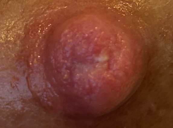
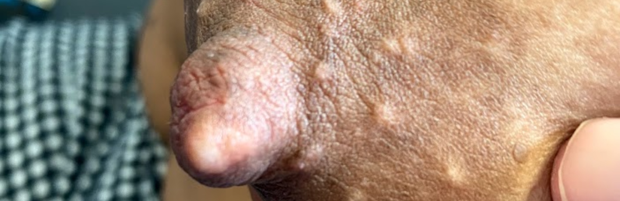
A hyperkeratotic spot of the nipple is an area of stratum corneum which has thickened in response to a focus of repetitive and excessively high mechanical trauma during breastfeeding or mechanical milk removal. A hyperkeratotic spot is often exquisitely painful in response to even mild pressure, perhaps because the thickened plaque of stratum corneum places pressure on the dermis, which is highly vascular and dense with sensory nerve endings.
A hyperkeratotic spot may appear pale white, cream, or yellowish, though colour alters during or immediately after milk removal due to the effects of moisture and epithelial hydration. A hyperkeratotic spot is larger than a milk blister, and with more defuse borders. Multiple, irregular sized hyperkeratotic spots may form on the face of a nipple which is subject to repetitive micro-trauma.
Attempts to unroof a hyperkeratotic spot, mistaking it for a milk blister, will worsen hyperkeratosis.
Hyperkeratotic white spots can be thought of as a lactation-related form of callous
When skin is rubbed or irritated repetitively over time, the stratified squamous keratinised epithelium thickens and the skin develops a patch of thickening to protect the area that is exposed to repetitive mechanical trauma. When this happens on the hands or feet, it is referred to as a callous. A callous is an even plaque of hyperkeratinised squamous epithelium. A corn has a central core of very dense keratin, surrounded by softer keratin.
Mostly, callouses are not painful, but sometimes, depending on their position, they can be acutely painful! Callouses don't usually have lymphocytes infiltrating them, in an inflammation, but this can happen particularly in corns.
Here is a photo of a hyperkeratotic spot before a breastfeed.
The O'Hara 2012 abstract
In 2012, a US medical practitioner Dr O’Hara published an abstract (without publishing an accompanying paper), which reported histological analysis of punch biopsies of painful white spots from five breastfeeding women.
-
The tissue was characterized as rubbery and scar-like.
-
Histological analysis was consistent with the diagnosis of hyperkeratosis:
-
No bacteria or fungi were identified
-
Leukocytes and fibrin were identified, signaling inflammation.
-
-
The women reported that their symptoms resolved shortly after biopsy removal.7
O’Hara reported that patients with white spots who subsequently presented to that clinic (number not stated) were effectively treated with a short daily course of mid-potency steroid under occlusion. A moist wound dressing (plastic wrap) was applied to enhance penetration of steroid into inflamed and fibrotic tissue. The author concluded that "nipple blebs are an inflammatory response to nipple trauma.…Clinicians should check for and treat any underlying causes of the recurrent trauma".7

Here is a photo of a hyperkeratotic spot after a breastfeed.
2. Milk blister (rare)
A milk blister is an exquisitely painful white spot or lesion on the nipple face, usually with a clearly demarcated border. It occurs rarely, and is sometimes associated with a lump or cord-like area extending from the nipple blister into the breast. Microscopic epithelial inflammation in the region of a duct orifice, most likely due to mechanical trauma, may heal so that the stratified squamous epithelium which extends two millimetres inside the orifice fuses during the inflammatory process of wound healing.
There is often a build-up of milk in the main duct behind the resultant milk blister, resulting in elevated intra-luminal pressure in the glandular tissue drained by branches of that duct. The latter triggers inflammation and high white cell counts,8 explaining the inspissated milk that is sometimes released or expressed when a milk blister is released.
3. Epidermal inclusion cyst (occasional)
Large epidermal inclusion cysts
This image shows what has been labelled by others as an epidermal inclusion cyst. Importantly, however, the boundaries of the lesion are irregular. Without examining this breastfeeding patient clinically, I wonder if this isn't more likely an image of severe hyperkeratosis, with inflammation of the surrounding tissues. A hyperkeratosis is not usually raised very much, but is a thickening of the epidermis.
Epidermal inclusion cysts are the most common cutaneous cyst in the human body. An epidermal inclusion cyst can also rarely occur on the nipple, not necessarily related to lactation, but may grow bigger in response to the mechanical pressures of lactation, and become inflamed and painful.
Epidermal inclusion cysts on the skin elsewhere in the human body are caused by implantation of epidermal elements into the dermis layer of the skin, with the cyst wall usually derived from the infundibular portion of the hair follicle.
An epidermal inclusion cyst contains keratinous material and cell debri, and can be conceptualised on a spectrum, of which a milium is the smallest. Although epidermal inclusion cysts elsewhere on the body are usually harmless and painfree, they may become sensitive on the nipple and even become so large and irritated that they require surgical removal so that breastfeeding can proceed comfortably.
Epidermal inclusion cysts don't contain sebum, but are often mislabelled as 'sebaceous cysts'.
Milium (occasional)
A milium is a painless, small white dermal cyst or epidermal inclusion cyst on the face of the nipple, or elsewhere on the nipple-areolar complex. A milium contains keratin, and is lined by a layer of stratified squamous epithelium. A milium cyst may appear prominent and very white after a breastfeed or mechanical milk removal, and less so otherwise. It usually disappears in time, and no treatment is required.
References
- Mitchell K, Eglash A, Bamberger E. Mammary dysbiosis and nipple blebs treated with intravenous daptomycin and dalbavancin. Journal of Human Lactation. 2020;36(2):365-368.
- Mitchell K, Johnson HM. Breast pathology that contributes to dysfunction of human lactation: a spotlight on nipple blebs. Journal of Mammary Gland Biology and Neoplasia. 2020:http://doi.org/10.1007/s10911-10020-09450-10917.
- Rodriguez JM, Fernandez L. Infectious mastitis during lactation: a mammary dysbiosis model. In: McGuire M, Bode L, editors. Prebiotics and probiotics in human milk: Academic Press; 2017. p. 401-428.
- Douglas P. Re-thinking benign inflammation of the lactating breast: a mechanobiological model. Women's Health. 2022;18:17455065221075907.
- Douglas PS. Re-thinking benign inflammation of the lactating breast: classification, prevention, and management. Women's Health. 2022;18:17455057221091349.
- Douglas PS. Re-thinking lactation-related nipple pain and damage. Women's Health. 2022;18:17455057221087865.
- O'Hara M. Bleb histology reveals inflammatory infiltrate that regresses with topic steroids: a case series. Breastfeeding Medicine. 2012;7 (Suppl 1):S2.
- Witkowska-Zimny M, Kaminska-El-Hassan E. Cells of human milk. Cellular and Molecular Biology Letters. 2017;22(11):doi:101186/s111658-101017-100042-101184.
- Mitchell KB, Johnson HM, Rodriguez JM, Eglash A, Scherzinger C, Cash KW, et al. Academy of Breastfeeding Medicine Clinical Protocol #36: The Mastitis Spectrum, Revised 2022. Breastfeeding Medicine. 2022;17(5):360-375.
- Douglas PS. Does the Academy of Breastfeeding Medicine Clinical Protocol #36 'The Mastitis Spectrum' promote overtreatment and risk worsened outcomes for breastfeeding families? Commentary. International Breastfeeding Journal. 2023;18:Article no. 51 https://doi.org/10.1186/s13006-13023-00588-13008.