
Your breasts' connective tissue not only holds everything together but helps regulate your milk glands
What is breast stroma made of?
Your milk glands and milk ducts are embedded in your breast's stroma, which includes connective tissue and fatty tissue. The amount of fatty tissue in your breast stroma decreases in pregnancy and during lactation, as the amount of your glandular tissue dramatically expands. Stroma also contains blood vessels, lymph vessels, neurons, cells which make collagen, a great abundance of immune cells, and also stem cells.
Your breasts - both the stroma and the glandular tissue - are defined by, and enclosed in, a fine layer of fascia - as if wrapped in muslin. This fascia runs superficially under the skin, then folds around deep behind the breast stroma to cover the front surface of your pectoral muscle. Your pectoral muscles sit snug against your ribcage.
A loosely defined framework of fibrous connective tissue is suspended between these superficial and deep fascial layers, in irregular dense bands. These fibrous bands support the breast’s glandular and fatty tissues and stabilise the breast. They naturally lengthen as the years pass.
What does breast stroma do when you're making milk?
Stroma is sensitive, highly bioactive tissue which not only provides structural support for your breast, but which interacts constantly with glandular and other cells in your breast to help regulate growth, immune responses, and gland function. This communication with the stroma happens
-
Through the myriad hormones and neurotransmitters released by cells in the stroma, and,
-
Very importantly, in response to mechanical pressures which are sensed within the stroma.
Understanding just how sensitive stroma is to mechanical pressure effects helps us understand how best to help if you develop a breast inflammation, such as mastitis or engorgement.
Recommended resources
Blood and lymph in your working breasts: an intertidal zone
The size of your breasts doesn't predict how much milk you'll make
Working breasts are diverse on the inside
Your glandular tissue
Working breasts are diverse on the outside
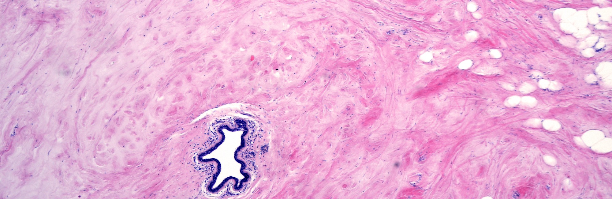
The photo at the top of this page shows a small lactiferous duct resting in the interlobular connective tissue or stroma of the breast. The duct is lined by columnar epithelial cells and is surrounded by myoepithelial cells.
Selected references
Bamberg EE, Maslanka M, Vinod-Paul K. Obesity-driven changes in breast tissue exhibit a pro-angiogenic extracellular matrix signature. Matrix Biology Plus. 2024;24(100162).
Ramsay DT, Kent JC, Hartmann RA, Hartmann PE. Anatomy of the lactating human breast redefined with ultrasound imaging. Journal of Anatomy. 2005;206:525-534.
Geddes DT, Aljazaf KM, Kent JC, Prime DK, Spatz DL, Garbin CP, et al. Blood flow characteristics of the human lactating breast. J Hum Lact. 2012;28(2):145–152. doi:10.1177/0890334411435414.